For more patient safety: 3D high-tech and robotics in the ENT OR at the MHH
The surgeons in the ENT operating theatre at Hanover Medical School are taking a double technical quantum leap: in future they will be operating with the latest generation of a fully digital and high-resolution 3D surgical microscope from Munich Surgical Imaging and a surgical robot for cochlear implantation. “The imaging with the 3D microscope is excellent. As a surgeon, you see the tiny structures much more precisely. Since we perform highly sensitive microsurgery, this new type of 3-D microscope is another leap forward in terms of patient safety,” reports ENT clinic director Prof. Prof. h.c. Dr. med Thomas Lenarz, who has established the world’s largest centre for cochlear implantation at the MHH.
But the new microscope also makes a valuable contribution to the residents’ further training in the operating theatre, because via the 3D glasses and the large monitors they see exactly what the surgeon sees through his binoculars and can follow every step in detail. Thus, during a cochlear implantation, they see the sensitive structures, which must not be accidentally damaged, in 3-D and can view the insertion of the electrode into the patient’s inner ear in unprecedented quality.
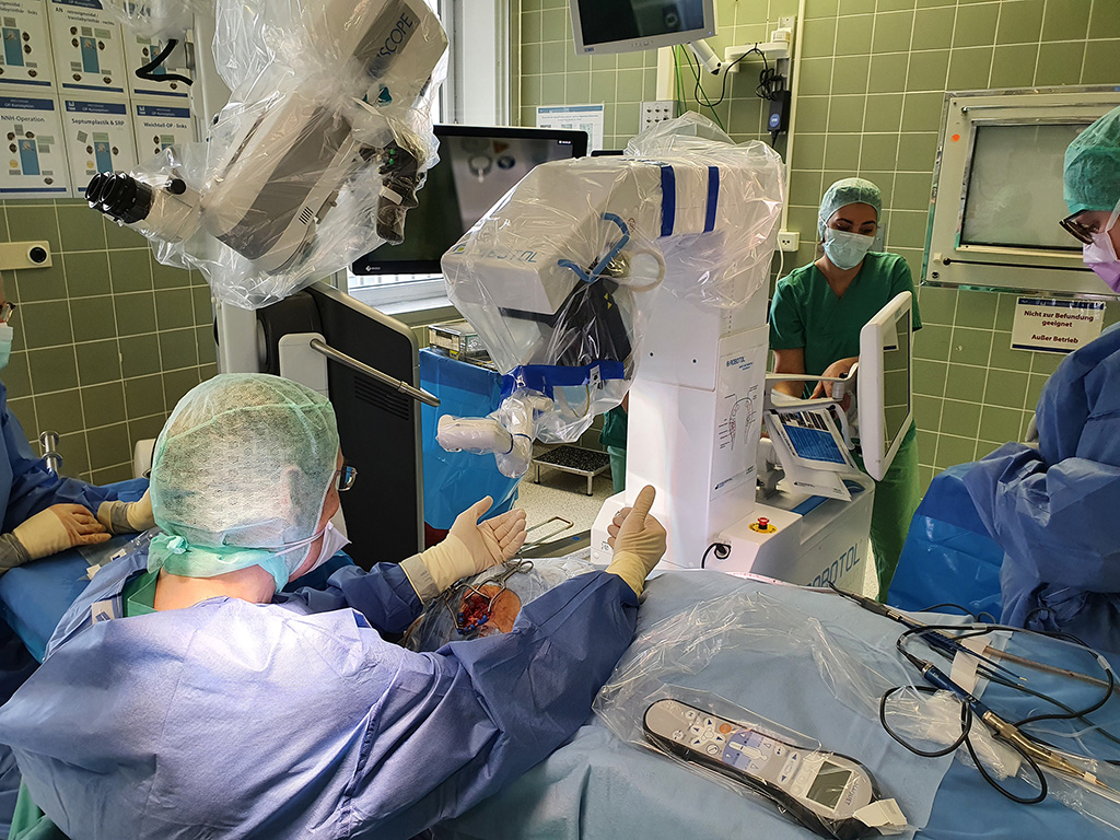
Another new addition is the “Robotol” surgical robot, which supports ENT surgeons in the sensitive phase of electrode insertion during cochlear implantation: The robot allows the precise insertion of the electrode into the cochlea along a pre-planned so-called trajectory, which is visualised in the planning computer tomogram. The insertion is therefore very slow and even – better than even experienced surgeons could do. This allows the sensitive structures of the cochlea to be protected. The ENT clinic of the MHH is the first clinic to have performed a robot-assisted cochlear implantation. Thus, precision medicine through robots is making its way into ear surgery.